Netter’s Atlas of Human Anatomy is a seminal work in medical education, renowned for its exquisite anatomical illustrations by Frank H. Netter and contributions from Carlos A.G. Machado. This 7th edition atlas is a cornerstone for medical students and professionals, offering detailed, clinically relevant depictions that bridge art and science in human anatomy.
1.1 Overview of the Atlas
Netter’s Atlas of Human Anatomy is a comprehensive guide offering detailed anatomical illustrations by Frank H. Netter and Carlos A.G. Machado; Designed for medical students and professionals, it provides a regional approach to anatomy, combining classic depictions with updated radiologic images and clinical correlations. The atlas is available in print and digital formats, ensuring accessibility for modern learners. Its clarity and precision make it an indispensable resource in medical education.
1.2 Importance in Medical Education
Netter’s Atlas is a cornerstone in medical education, providing detailed, clinically relevant illustrations that aid students and professionals in understanding human anatomy. Its precise depictions and regional approach make it an essential tool for learning and reference. As the gold standard, it bridges art and science, offering unparalleled clarity and accuracy for medical training and practice.
History and Evolution of the Atlas
First published in 1989, Netter’s Atlas has evolved through editions, incorporating contributions from Frank H. Netter and Carlos A.G. Machado, with updated content and digital features.
2.1 Frank H. Netter and His Contribution
Frank H. Netter, MD, is celebrated as one of the most influential medical illustrators in history. Known as the “Medical Michelangelo,” his detailed, accurate, and artistically superior anatomical illustrations laid the foundation for the atlas. His work emphasizes clarity and clinical relevance, making it indispensable for medical education and practice. Netter’s legacy continues to inspire, with his illustrations remaining the gold standard in anatomical representation.
2.2 Editions and Updates Over the Years
First published in 1989, Netter’s Atlas has undergone significant updates, with the 7th and 8th editions introducing new radiologic images, clinical tables, and contributions from Carlos A.G. Machado. Each edition enhances anatomical accuracy and clinical relevance, ensuring the atlas remains a vital resource. Digital versions now offer interactive tools, expanding its utility for modern learners and professionals alike.

Key Features of the Netter Atlas
Netter’s Atlas features detailed anatomical illustrations, clinical correlations, and interactive digital versions, providing comprehensive anatomical views and modern tools for medical education and professional reference.
3.1 Detailed Anatomical Illustrations
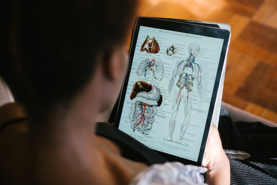
The atlas is celebrated for its precise and visually stunning illustrations, created by Frank H. Netter and updated by Carlos A.G. Machado. These detailed depictions provide clear, lifelike representations of human anatomy, emphasizing clinical relevance and anatomic relationships. The artwork is renowned for its clarity and accuracy, making complex structures easily comprehensible for both students and professionals in the medical field.
3.2 Clinical and Radiological Correlations
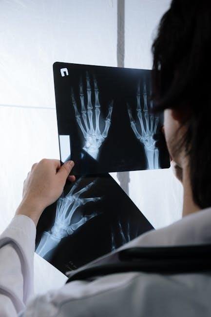
The atlas seamlessly integrates anatomical illustrations with clinical and radiological images, enhancing understanding of real-world applications. This correlation aids in diagnosing and surgical planning, offering a holistic view of human anatomy. The inclusion of radiologic images bridges the gap between theoretical knowledge and practical medicine, making it an invaluable resource for both students and practicing professionals.
3.4 Interactive and Digital Versions
The Netter Atlas is available in interactive digital formats, offering enhanced learning tools such as 3D models, videos, and multiple-choice questions. These features allow users to explore anatomy dynamically, trace pathways, and reinforce clinical concepts. The digital versions provide flexible access, enabling students and professionals to study efficiently and engage with anatomical content in innovative ways, complementing the traditional print edition.
Structure and Organization
Netter’s Atlas is organized regionally, providing comprehensive coverage of the human body. It systematically presents anatomical structures, facilitating a clear understanding of complex systems and their relationships.
4.1 Regional Approach to Anatomy
Netter’s Atlas employs a regional approach, organizing anatomy by body regions to enhance understanding of spatial relationships. This method mirrors dissection and clinical practice, making it ideal for medical students. Each region is meticulously illustrated, with detailed depictions of structures and their interconnections. The atlas also integrates clinical correlations, providing real-world context for anatomical knowledge. This approach ensures a logical and practical learning experience, bridging the gap between theory and application.
4.2 Comprehensive Coverage of Body Systems
Netter’s Atlas provides an extensive exploration of all major body systems, ensuring a thorough understanding of human anatomy. From the skeletal and muscular systems to the nervous and circulatory, each system is detailed with precise illustrations. The atlas also includes radiological correlations, offering a modern and integrated perspective. This comprehensive approach makes it an invaluable resource for both students and professionals in the medical field, fostering a deep appreciation of anatomical complexity and clinical relevance. The detailed coverage ensures that learners gain a holistic view of the human body, aiding in diagnosis and treatment. Additionally, the inclusion of clinical tables and updated radiologic images enhances the atlas’s utility in contemporary medical education and practice.

The Artistry Behind the Atlas
Frank H. Netter’s remarkable artistic skill and attention to detail have made his illustrations iconic in medical education. His work, often called “Medical Michelangelo,” combines precision with beauty, creating vivid, intricate depictions of human anatomy that are both informative and aesthetically pleasing, setting the atlas apart as a masterpiece in the field.
5.1 Frank H. Netter’s Artistic Legacy
Frank H. Netter’s artistic legacy is unparalleled in medical illustration. His meticulous, lifelike depictions of human anatomy, often described as the work of the “Medical Michelangelo,” have set a standard for clarity and beauty. Combining scientific precision with artistic flair, Netter’s illustrations have educated generations of healthcare professionals, making his work indispensable in medical education and practice worldwide.
5.2 Modern Contributions by Carlos A.G. Machado
Carlos A.G. Machado has significantly enhanced Netter’s legacy with his contemporary medical illustrations. His detailed, anatomically precise artwork in the 7th edition seamlessly blends tradition with modern techniques, offering updated perspectives and radiologic correlations. Machado’s contributions ensure the atlas remains a vital resource for medical education, bridging the gap between classic and cutting-edge anatomical knowledge for students and professionals alike.

Educational Impact and Reception
Netter’s Atlas of Human Anatomy is the world’s best-selling anatomy atlas, celebrated for its clarity and clinical relevance. It is a favorite among medical students and professionals, offering detailed illustrations and updated content that make it an essential resource for anatomy education.
6.1 Popularity Among Medical Students
Netter’s Atlas of Human Anatomy is a favorite among medical students for its clear, detailed illustrations and clinical relevance. The 7th and 8th editions, featuring contributions from Frank H. Netter and Carlos A.G. Machado, are praised for their accuracy and user-friendly design. Students appreciate the atlas’s ability to simplify complex anatomical concepts, making it an indispensable tool for study and exam preparation.
6.2 Reviews and Recommendations by Professionals
Professionals widely endorse Netter’s Atlas for its precise, clinically relevant illustrations. Often described as the “gold standard” in anatomy, it is praised by educators and practitioners alike. The 7th and 8th editions are particularly commended for their updated radiologic images and integrated clinical tables, enhancing its utility in both surgical planning and diagnostic imaging. Many consider it an essential resource for modern medical practice.
Availability and Access
Netter’s Atlas is widely available in PDF and eBook formats, accessible via platforms like Scribd and Google Drive. Purchase options include digital and print editions.
7.1 PDF Versions and Digital Accessibility
The Netter Atlas of Human Anatomy is widely available in PDF format, accessible via platforms like Scribd and Google Drive. Digital versions offer enhanced accessibility, enabling users to download and view the atlas on various devices. The PDF format allows for easy searching, portability, and integration with modern study tools, making it a popular choice for medical students and professionals worldwide.
7.2 Purchase Options and Editions
The Netter Atlas of Human Anatomy is available in multiple editions, including the 7th and 8th editions, each offering updated content and illustrations. Purchase options include hardcover and eBook versions, accessible through major retailers and Elsevier’s official website. The Professional Edition caters to experts, while student editions provide affordable access to essential anatomical knowledge, ensuring versatility for diverse learning needs.
Comparisons with Other Anatomy Atlases
Netter’s Atlas stands out for its artistic precision and clinical relevance, surpassing competitors like Gray’s Anatomy in visual clarity and anatomical detail, making it a preferred choice among medical professionals and students alike.
8.1 Unique Selling Points
Netter’s Atlas of Human Anatomy is celebrated for its artistic precision and clinical relevance, offering detailed, hand-painted illustrations by Frank H. Netter and Carlos A.G. Machado. Its 7th edition includes updated radiologic images and clinical tables, enhancing its utility for both students and professionals. The atlas also provides an eBook version, making it accessible and user-friendly for modern learners.
Its unique blend of traditional artwork and digital accessibility sets it apart from competitors, ensuring comprehensive anatomical understanding while maintaining a clinician’s perspective. This dual approach solidifies its position as a gold standard in medical education and practice.
8;2 Competing Atlases and Their Features
Competing atlases, such as Gray’s Anatomy and Grant’s Atlas, offer detailed anatomical representations. Gray’s emphasizes comprehensive text and images, while Grant’s focuses on a regional approach. Sobotta’s Atlas provides cadaveric dissections, appealing to surgical and clinical needs. Each has unique strengths, but Netter’s artistic clarity and clinical correlations remain unrivaled, making it the preferred choice for many professionals and students.
While alternatives exist, Netter’s Atlas stands out for its precise illustrations and user-friendly design, ensuring its status as a gold standard in anatomy education and practice.

Clinical Applications
Netter’s Atlas aids in surgical planning and diagnostic imaging, providing clear anatomical insights. Its detailed illustrations help clinicians visualize complex structures, enhancing precision in medical procedures and education.
9.1 Use in Surgical Planning
Netter’s Atlas is a valuable resource for surgeons, offering detailed anatomical illustrations that aid in preoperative planning. Its clear, precise depictions of human anatomy enable clinicians to visualize complex structures, enhancing surgical accuracy and patient outcomes. The atlas’s clinically relevant artwork helps in understanding spatial relationships, making it an indispensable tool for surgical preparation and intraoperative decision-making.
9.2 Diagnostic Imaging Integration
Netter’s Atlas seamlessly integrates with diagnostic imaging, enhancing the correlation between anatomical illustrations and radiological findings. This integration aids clinicians in interpreting MRI, CT scans, and X-rays by providing a visual reference for complex structures. The atlas’s detailed artwork complements imaging modalities, offering a comprehensive understanding that improves diagnostic accuracy and informs treatment plans effectively.
Supplements and Additional Resources
Netter’s Atlas offers additional resources like eBooks, interactive modules, and study aids, enhancing learning and practical application of anatomical knowledge through diverse multimedia tools.
10.1 Companion Websites and Tools
Companion websites and tools enhance the Netter Atlas experience, offering interactive features like 3D models, videos, and multiple-choice questions. These resources provide hands-on learning opportunities, allowing users to explore anatomy dynamically. The interactive dissection modules and labeled/unlabeled figure versions aid in self-study and exam preparation. Additionally, access to an eBook version ensures flexibility and convenience for learners on the go.
10.2 Study Aids and Interactive Modules
Study aids and interactive modules complement the Netter Atlas, offering 300 multiple-choice questions, videos, and 3D models. These tools allow learners to trace pathways, test spatial knowledge, and engage with clinical concepts. Interactive plates enable self-assessment, while the modular design supports focused learning. These resources enhance understanding and retention of complex anatomical information for both students and professionals.
The Role of the Atlas in Modern Medicine
Netter’s Atlas remains a cornerstone in medical education and practice, bridging art and science with detailed, clinically relevant illustrations. Its digital versions enhance accessibility, supporting modern learning and clinical applications.
11.1 Bridging Art and Science
Netter’s Atlas seamlessly merges artistic brilliance with scientific precision, offering detailed anatomical illustrations that enhance understanding. Frank H. Netter’s medical illustrations, complemented by Carlos A;G. Machado’s modern contributions, create a visually engaging and educationally rich resource. This integration of art and science makes the atlas indispensable for medical students and professionals, balancing aesthetics with functional learning.
11.2 Future Developments and Innovations
Future editions of Netter’s Atlas are expected to integrate advanced technologies, such as 3D modeling and interactive digital platforms, enhancing the learning experience. The inclusion of new imaging techniques and updated clinical correlations will ensure the atlas remains a cutting-edge resource for medical education and practice, continuing its legacy as a leader in anatomical studies.

User Feedback and Testimonials
Users praise Netter’s Atlas for its clarity and detailed illustrations, making it a go-to resource for both students and professionals. Testimonials highlight its effectiveness in bridging art and science, with many appreciating the integration of clinical correlations and modern digital enhancements that enhance learning and practice.
12.1 Student Experiences
Medical students worldwide acclaim Netter’s Atlas for its clear, detailed illustrations and clinical correlations, making complex anatomy accessible. Many highlight its role in exam preparation and ease of understanding, while others appreciate the user-friendly format and integration of modern tools, fostering a deeper grasp of human anatomy and its practical applications in medicine.
12.2 Professional Endorsements
Professionals widely endorse Netter’s Atlas for its exceptional clarity and clinical relevance. Many highlight its detailed illustrations and practical correlations, making it indispensable in medical education and practice; Its status as the “gold standard” in anatomy resources is underscored by its popularity among educators and clinicians, who praise its ability to simplify complex anatomical concepts while maintaining precision and aesthetic appeal.
Netter’s Atlas of Human Anatomy remains a cornerstone in medical education, blending art and science. Its detailed illustrations and updates ensure continued relevance, solidifying its legacy as the gold standard for anatomical study.
13.1 Legacy and Continued Relevance
Frank H. Netter’s Atlas of Human Anatomy has left an indelible mark on medical education, celebrated for its artistic precision and scientific accuracy. With contributions from Carlos A.G. Machado, the 7th edition maintains its relevance, offering updated illustrations and radiologic correlations. Its availability in digital formats ensures it remains a vital resource for modern learners, solidifying its legacy as a foundational anatomical guide.
13.2 Final Thoughts on the Atlas’s Impact
Frank H. Netter’s Atlas of Human Anatomy remains a cornerstone in medical education, blending art and science seamlessly. Its detailed illustrations and clinical correlations have shaped generations of healthcare professionals. With updates like the 7th edition and digital accessibility, the atlas continues to inspire and educate, ensuring its lasting impact on the field of anatomy and beyond.
